
Model for virtual vascular reconstruction in liver transplantation using computational fluid dynamics
Boris Yaremin1, Bakhtiyar Kazymov1, Kamran Alekberov1, Andrey Russkikh2, Ramiza Sharifova3, Murad Novruzbekov1.
1Liver Transplantation Center, Sklifosovsky Emergency Medicine Institute, Moscow, Russian Federation; 2Operative Surgery And Topographic Anatomy Chair, Krasnoyarsk State Medical University, Krasnoyarsk, Russian Federation; 3Transplantation Scientific Group, Reaviz Medical University, Moscow, Russian Federation
Introduction: Computational anatomy is a modern research method that has become available using computer technology and computational fluid dynamics. It is promising to use it for perioperative planning and modeling, including organ transplantation. Aim of study is to prepare investigational model of blood flow in liver during liver transplantation.
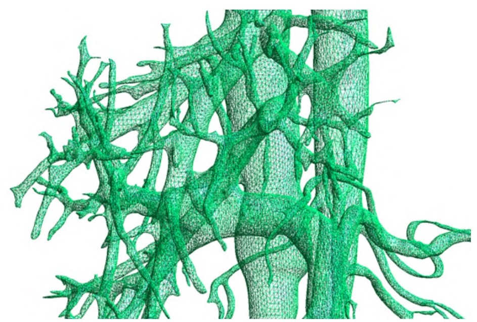
Matherials and methods: Contrast research was done among 53 men who were patients in surgery departments at Krasnoyarsk regional hospital #1, Russia (KRH). Estimations were made using 3D models of portal system (working stations GE Advantage Workstation, Siemens singo.via), based on multi-slices computed tomography of abdominal cavity with bolus contrasting with help of medication “Ultravist-370”. The volume of the used contrasting stuff made 100 ml, the speed of infiltration was 4 ml per second, and the average radial strain was 11,3 mЗv. DICOM data segmentation was performed using Dragonfly software (Object Research Systems, Canada) at the Innovation Technology Management Resources of the Reaviz University. Using series with arterial and vein contrast, we performed the segmentation of contrasting vessels, and obtained three-dimensional data topology in .obj format. Then we processed the obtained models using scripts prepared for the pythonOCC framework. We built the central lines of the vessels and formed the branching tree. Methods of computational hemodynamics were implemented using the Visual-CFD application for OpenFOAM environment (ESI, France). Mesh example of portal vein shown in figure 1.
Results: With the use of created three-dimensional computer model, the blood flow in the portal vein and liver arteries at various variants of its structure was simulated. It was obtained that, in the presence of the main type of structure with predominance of blood flow along the splenic vein, the blood flow turbulence and risk of thrombosis development are higher. At the same time, with virtual thrombosis of the portal vein trunk, the pressure gradient is 1.4 times higher than with the bulk type, which is more favorable for the proposed reconstruction. Thus, these data can be used for preoperative planning in surgical treatment of portal vein thrombosis in liver transplantation.
Conclusion: The proposed model of blood flow is promising and allows you to create an interactive tool for calculating the hemodynamic situation in various reconstructions during liver transplantation.

right-click to download