
Impact of autophagy on the development of senescence in protocol biopsies in kidney transplant patients
Maria C Giordani1, Carolina Vecino2, Silvia B Cristianssen3, Alejandro Ropolo2, Susana K Villamil cortez1, Nora c imperiali1, Silvia R Groppa1, Maria I Vaccaro2.
1Nephrology Department, Transplant Division, Hospital Italiano de Buenos Aires, Buenos Aires, Argentina; 2Department of Pathophysiology, Institute of Biochemistry and Molecular Medicine (UBA-CONICET), Schoo, University of Buenos Aires, Buenos Aires, Argentina. Conicet, Buenos Aires, Argentina; 3Pathology, Hospital Italiano de Buenos Aires, Buenos Aires, Argentina
Introduction: In kidney transplant, the expression of cyclin-dependent kinase inhibitors p16INK4a, a cellular senescence marker, is a strong predictor of poor allograft outcome. Autophagy is a tightly regulated cellular self-degradation recycling process characterized by sequestration of damage organelles in double-membrane vesicles called autophagosomes, as a cellular survival mechanism under stress. VMP1 vacuole membrane protein1 is a transmembrane protein involved in the initial steps of the autophagy process. Patients with chronic allograft dysfunction have downregulation of autophagy. Both senescence and autophagy are activated in response to kidney injury during transplantation. A relationship between both seems to exist, although it is still poorly understood, and human studies are scarce. A better understanding of this phenomena could allow the development of therapies to increase graft longevity. We aim to assess the relationship between senescence and autophagy through tissue expression of p16INK4a and VMP1 in renal transplants patients.
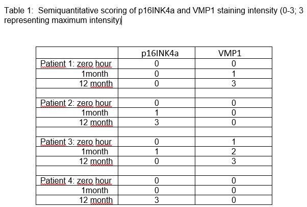
Materials and Methods: Protocol transplant(Tx) kidney biopsies at zero hour, 1 and 12 post Tx months in patients who consent were included. Immunohistochemistry staining for p16 INK4a was performed on paraffin biopsy sections with peroxidase anti-peroxidase Ventana BENCHMARK- XT. Immunofluorescence with antibodies to Polyclonal rabbit antiserum to VMP1 were used. We evaluated the extent of nuclear p16INK4a staining and VMP1 positive expression as a punctate cytoplasmic structure in the renal cortex. Semiquantitative scoring of p16INK4a and VMP1 staining intensity (1-3; 3 representing maximum intensity) was analyzed.
Results: 12 protocol biopsies from 4 cadaveric kidney tx patients were analyzed. All patients had triple immunosuppressive regimen and none had rejection in the biopsies. VMP1 was negative in glomeruli (podocyte, endothelial and mesangial cell) in all biopsies. In VMP1 positive samples, it was found as granular intracytoplasmic vacuole in proximal and distal tubule cells and in arterial smooth muscle cells. Nuclear p16INK4a staining was positive in proximal tubules and glomerular endothelial cells. Semiquantitative analysis of the expression of both markers is represented in Table 1. A progressive increase in the expression of the autophagy marker VMP1, from the pre-implantation biopsy to the year after transplantation, was seen in samples from patients (1 and 3) who had no expression of the senescence marker p16 INK4a. Opposite to this, patients 2 and 4 expressed p16 INK4 at one year after transplantation and had no expression of VMP1.
Conclusion: In renal biopsies from 4 kidney Tx patients, those who expressed the cell-protective autophagy marker VMP1 did not express the senescence marker p16 INK4a.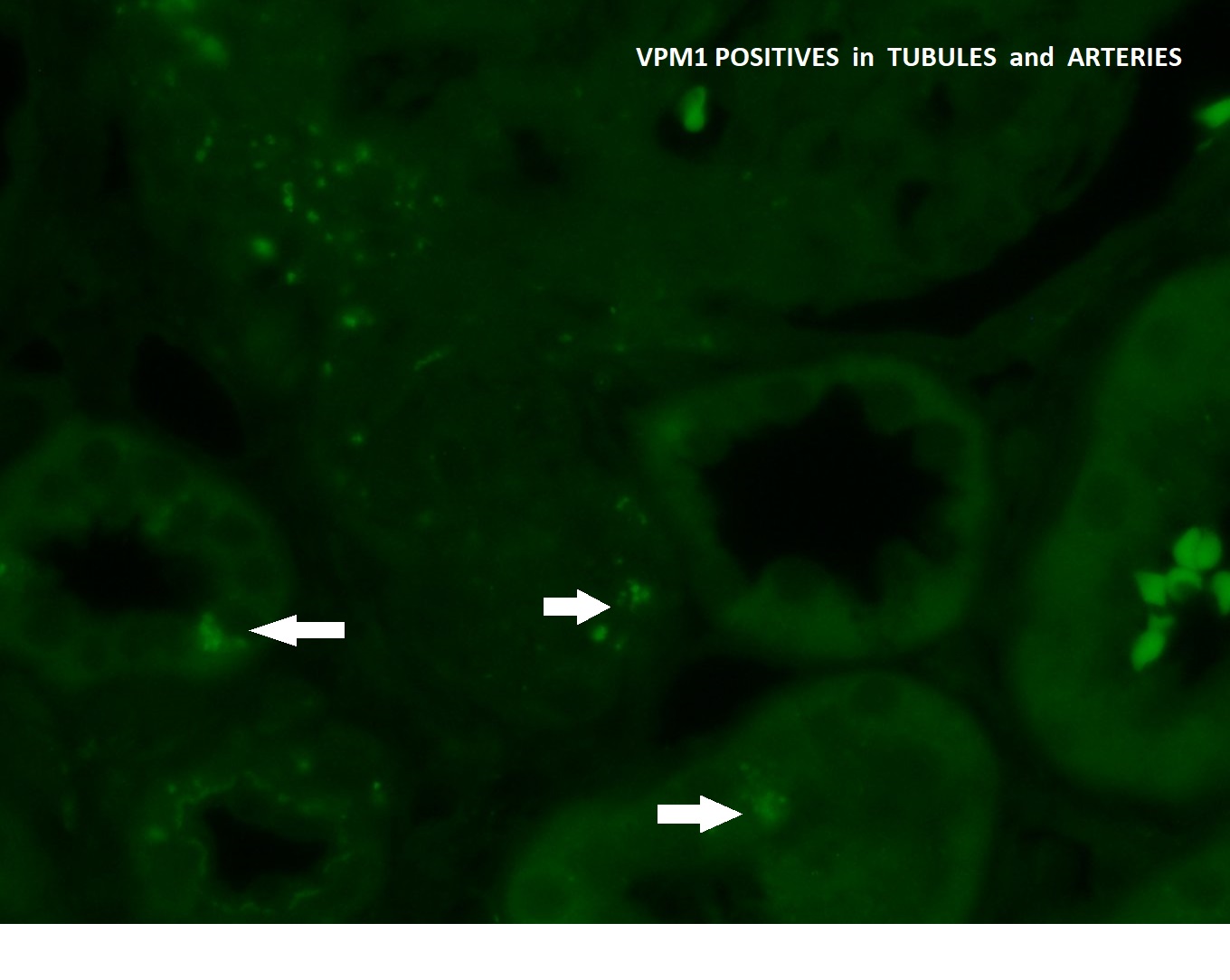

right-click to download