

In vivo monitoring of liver organoids differentiation using live-screening-optical technologies
Giuseppe Pettinato1, Mark F. Coughlan1, Xuejun Zhang1, Liming Chen1, Maria Glyavina1, Yuri N. Zakharov1, Lei Zhang1, Le Qiu1, Robert A. Fisher2, Lev T. Perelman1.
1Department of Medicine, Center for Advanced Biomedical Imaging and Photonics, BIDMC, HMS, Boston, MA, United States; 2CTI Clinical Trial & Consulting, CTI, Midlothian, VA, United States
Introduction: Tissue engineering is a promising technology for the cure of liver diseases 1-2. Engineered liver organoids have been demonstrated to be a potential alternative to isolated liver primary hepatocytes 3-5. Monitoring the progression of the differentiation process in live cells has great implications for the obtainment of functionally differentiated liver organoids. In this study, we have applied live-screening-optical technologies to monitor the progression of our differentiated human induced pluripotent stem cells (hiPSCs) in order to be able to improve the maturation and function of our liver organoids by looking at live chromatin remodeling and various other liver markers 6-7.
Methods: The generation of human embryoid bodies (hEBs) was achieved utilizing HLA-Class II knockout hiPSCs mixed with endothelial cells, employing our ROCKi/Spin-free technology 8. Using confocal light scattering spectroscopic microscopy technology we monitored the chromatin remodeling of our hEBs during the whole differentiation process. This analysis allowed us to characterize our four-stage hepatocyte differentiation protocol according to the whole genome modifications observed. We also carried out classical biochemical analyses to confirm results obtained with our live-screening-optical technologies.
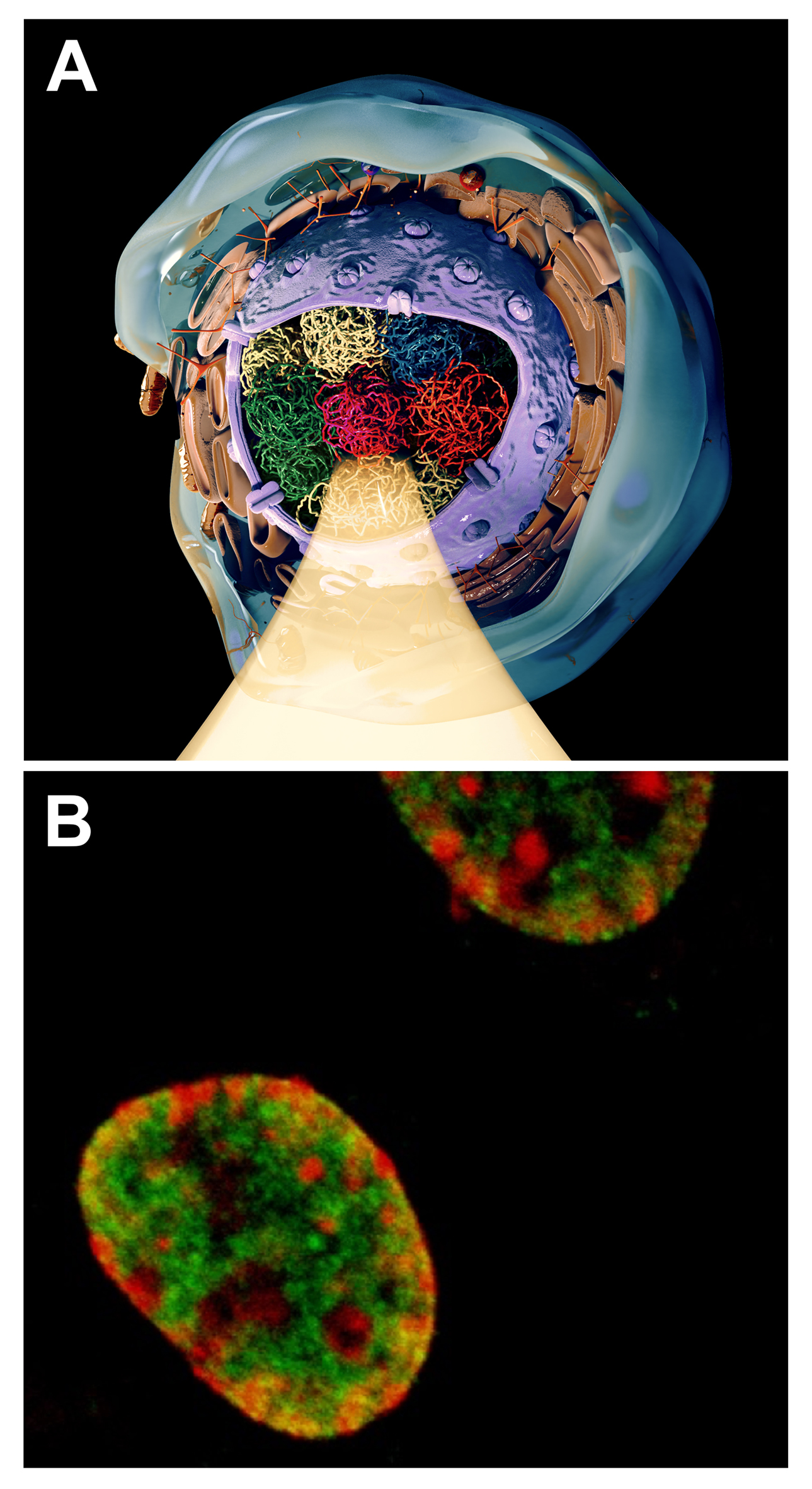
Results: Differentiating liver organoids were followed by performing real-time chromatin remodeling to show changes in chromatin structure (euchromatin vs heterochromatin) over the course of the differentiation process (Fig. 1 A and B). Gene expression of several hepatic markers, as well as coagulation factors, were detected by real-time PCR showing a progression in the maturation of our liver organoids.
Conclusions: Real-time monitoring of our differentiating liver organoids represents a great tool to evaluate the differentiation advancement in live cells. Our data demonstrated the ability of confocal light scattering spectroscopic microscopy to provide critical information of the differentiation status in vivo during liver differentiation, providing a new tool necessary for the progression to clinical translation.
References:
1. Takahashi, K. et al. Cell 131, 861-872 (2007).
2. Yu, J. et al. Science (New York, N.Y.) 318,1917-1920 (2007).
3. Si-Tayeb, K. et al. Hepatology (Baltimore, Md.) 51, 297-305 (2010).
4. Pettinato G, et al. Sci Rep. 2016 Sep 12;6:32888.
5. Pettinato G, et al. Sci Rep. 2019 Jun 20;9(1):8920.
6. Keenen B, et al. J Cell Physiol. 2009 Apr;219(1):1-7.
7. Gökbuget D, et al. Development. 2019 Sep 25;146(19). pii: dev164772.
8. Pettinato, G., et al. Sci Rep 4, 7402 (2014).

right-click to download